Ocular Surface and Translational Research Lab
The Lab runs the dual role of "bench and bedside", carrying out research on tissues and fluids of the eye-- in particular tears, conjunctival and corneal cells, secretion of meibomian cells, cell cultures and in vitro organ cultures. The in vivo research is conducted in an area for the evaluation of outpatients, with specific instrumentation for the ocular surface.
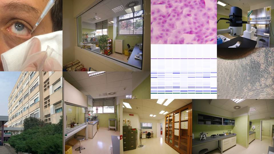
The Lab, designed in accordance with the appropriate legislation, covers a total area of 200 sq m, divided into different activity areas. The diagnostic activity concerns samples from human subjects, collected in the dedicated outpatient collection area.
The diagnostic and research activity is focused on specific samples (tears, epithelial cells of the cornea and conjunctiva, secretion of the meibomian glands), which can only be taken in minute quantities, insufficient for the automated instruments of centralized laboratories.
Research fields:
Pathophysiology of the ocular surface, medical substances (tear substitutes) for the treatment of ocular surface diseases, blood components for non-transfusion use (EUNT) for the biological treatment of severe keratopathies, and slow-release biomaterials (drugs and growth factors). Neuroprotection mechanisms using in vitro models.
More details on the dedicated page.
Teaching activities:
The Lab is attended by Alma Mater students for practical internships and preparation of the degree or doctoral thesis, specifically: students of the School of Sciences [Degree in Biological Sciences], School of Pharmacy [Master's Degree in Health Biology, Biosanitary-Forensic and Nutritional courses], School of Medicine and Surgery [Degree of Medicine and Surgery, Techniques of Biomedical Laboratory, School of Specialization in Ophthalmology].
Highly specialized courses are organized in collaboration with the Alma Mater Foundation, accredited for Continuing Medical Education, and aimed at various professionals operating in the health sector.
Equipment by area:
-
Cell culture area with double entry/exit: air filtration system, air conditioning, controlled relative humidity and carbon dioxide concentration, equipped with incubators for cell cultures (at temperature and CO2), refrigerator for storage of reagents, laminar flow cabinet for processing cells and tissues in a controlled environment (class A according to Annex 1 cGMP), centrifuges and microcentrifuges, thermostatic bath, dissection microscope (Stemi 2000 KL750 ZEISS), inverted microscope equipped with fluorescence (Axiovert 10 ZEISS).
-
Molecular biology area: Thermal Cycler 2720 Applied Biosystems, Qbit 2.0 fluorometer Invitrogen.
-
Preparation, histology and staining area:chemical hood, refrigerated centrifuge, microtome, thermostatic bath for histology, dry ovens, analytical balance, ZEISS optical microscopes equipped with fluorescence and phase contrast, safety cabinet for reagents.
-
Protein analysis area: BIO-RAD 1D electrophoresis cells, Thermo Scientific Multiskan Sky Touch Screen ELISA reader, Agilent Bioanalyzer 2100.
-
Cold zone with + 4 ° C refrigerators, -20 ° C and -80 ° C freezers, cryogenic containers.
-
Patient study area: LED slit lamp equipped with image acquisition system, infrared lamp and cobalt blue filters, data storing software. Equipment for infrared meibography, meniscometry, non-invasive tear break-up time, blink analysis, ocular protection index (OPI), pupillometry (Topcon CA 800), meibometry (Courage & Khazaka), TearLab Osmometer.
Responsible
-
Associate Professor
Dipartimento di Scienze Mediche e Chirurgiche - DIMEC
Via Massarenti 9
Bologna (BO)
Tel: +39 051 2142850
Fax: +39 051 636 2 846
Team
Chiara Coslovi
Area dei Collaboratori - Settore socio-sanitario
Collaborators:
Claudia Pancani – TdLB – AOSP
Gloria Astolfi- University of Bologna
The Laboratory hosts colleagues from collaborating research groups.